 |
 |
|
|
|
St. John's wort has long been used for different medicinal purposes. The plants grow spontaneously in nature. Because of great phytopharmaceutical need St. John's wort in many countries has commercially been grown over the last few years. Initial endeavors to produce St. John's wort in plantations in Serbia were unsuccessful. All grown plants were affected by diseases and destroyed.
The aim of this paper is to find the cause of St. John's wort wilting and root and crown rot in Serbia. Eight strains of Fusarium oxysporum were isolated from diseased roots, crown and stems of St. John's wort. Pathogenecity test showed that four were pathogenic to St. John's wort plants. Diseases symptom, characteristic for natural infection, was also reproduced by artificial inoculation of St. John's wort plants in greenhouse. In addition to morphology studies of the isolated fungi effect of different media, temperature and pH of media on colony growth were investigated.
St. John's wort (Hypericum perforatum L.) has so far not been commercially grown in Serbia. But, because of its increasing use as antidepressant medication there is great interest for its commercial growing. At the Institute for Medicinal Plant Research "Dr Josif Pančić", Belgrade, St. John's wort growing has been initiated from 1997-1999 (Radanović et al., 2000).
The plants were grown on heavy soil (black marsh soil), with 48.6% of clay 45.9% powder 5.5% sand with average content of humus (3.5%). In investigation is used local population of St. John's wort from western Serbia origin.
In 1999, there were four St. John's wort plantations in experimental field in Pančevo. The two years plantations were planted in fall 1997 and spring 1998, and one year plantations were planted in fall 1998 and spring 1999. In 1999 growth retardation, wilt and completely plants death, resembling symptoms of Fusarium wilt have been observed. The disease has spread steadily to all plants, so, after first flowers harvesting all plants in all of four plantations have died.
Fusarium oxysporum is causing many similar diseases on medicinal plants like that one on basil in Italy (Tamietti and Matta, 1989) and recently in Israel (Gamliel et al., 1996). So far there are 64 different formae speciales of F. oxysporum registered on different plants (Watanabe, 1994). Besides Srebia, Fusarium will, crown and root of St. John's wort is a problem in Bulgaria as well (Evstantieva, 2000 (personal communication). While Fusarium diseases of St. John's wort is not a problem in Canada [Branka Barl, 2000 (personal communication)]. The aim of this paper was to find the cause of St. John's wort wilting and root and crown rot in Serbia.
Isolation of culture
Plants collected in the field with symptoms on roots, crown and stems were thoroughly washed free of soil particles and later rinsed in sterile distilled water. Segments of 2 cm long were taken from root and along the stems of diseased plants, dipped in 70% ethanol, surface-sterilized with NaOCl (1%) for 1 min., and rinsed in sterile water. Each segment was then cut into three pieces, which were blotted and placed on potato-dextrose agar (PDA) (Dingra and Sinclair, 1995). Plates were incubated in the dark at 27°C for 3-7 days, and colony of F. oxysporum were identified microscopically (Watanabe, 1994).
Pathogenecity test
Local populations of ten-week-old St.John's wort plant, growing in potting mix. in 10 cm pots, were inoculated by injecting 10 ml of suspension of conidia 5-10 cm from both side of root zone. Plants were grown in a greenhouse at 90% relative humidity and 25 to 35°C.
No inoculated St. John's wort plants served as control and were maintained under the same conditions. The non-inoculated plants showed no disease symptoms.
Colony growth
The effect of temperature, media, pH and light on colony growth was studied in separate experiments. Discs of 5 mm diameter of 5 days old colony was transferred to the center of Petri plates with nutrient media. The colony diameter was measured every second day until the colony almost field 9 cm Petri plates. Effect of temperature, pH was studied on PDA and the effect of different media was studied on: PDA, water agar (WA), corn meal agar (CMA), oatmeal agar (OMA), Czapec agar (CA) and maltz-extract agar (MA) media.
Identification of the pathogen was done by morphology study of mycelia, microconidia, macroconidia and hlamidospores and by colony growth on different media and temperatures.
Symptoms
The first sign of disease in 1999, appeared when the plants were 10-15
cm. Some plants died very young and some after flowering. The disease starts
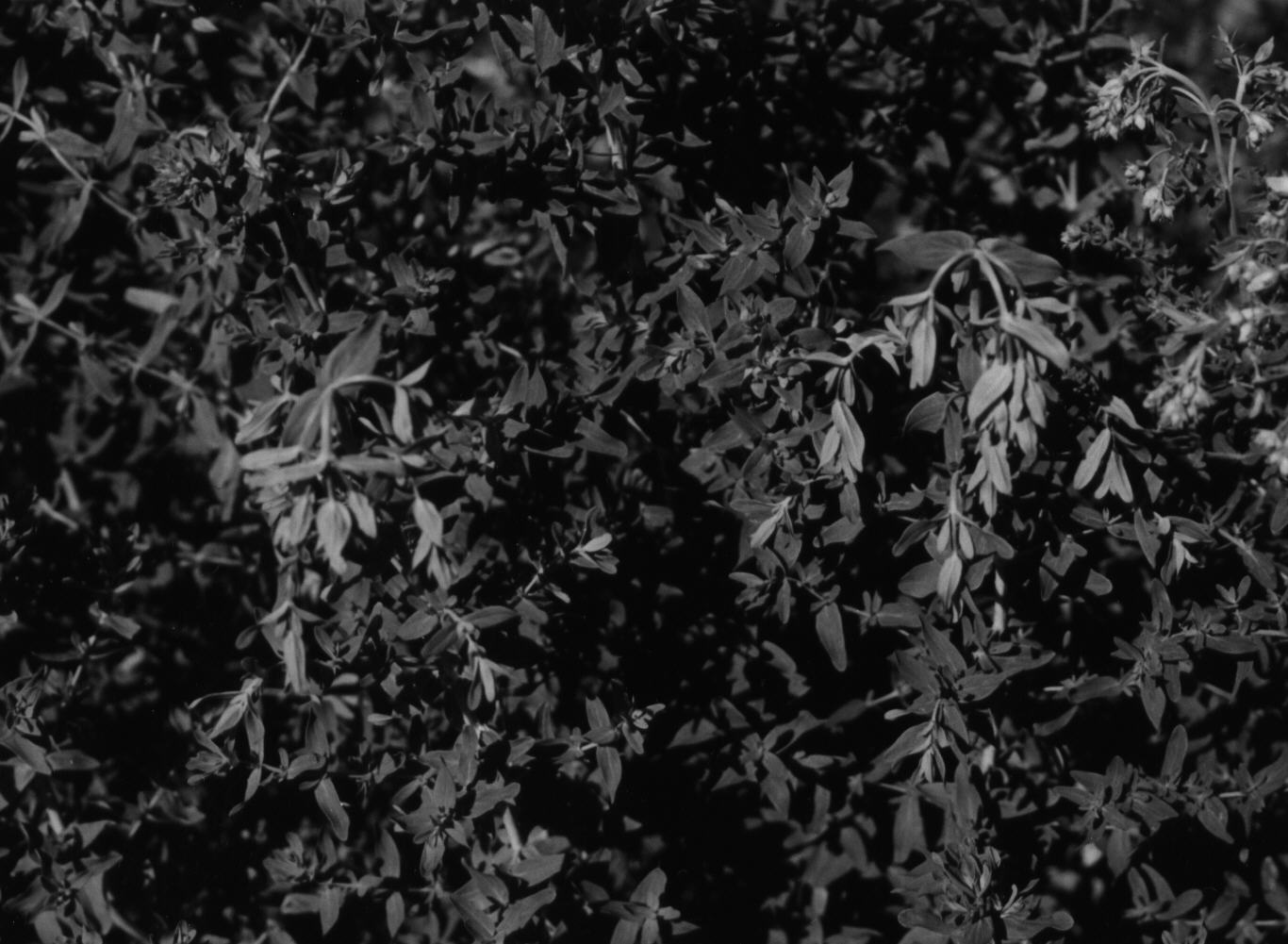
with wilting of the top of plant (fig. 1), yellowing and necrosis of leaves
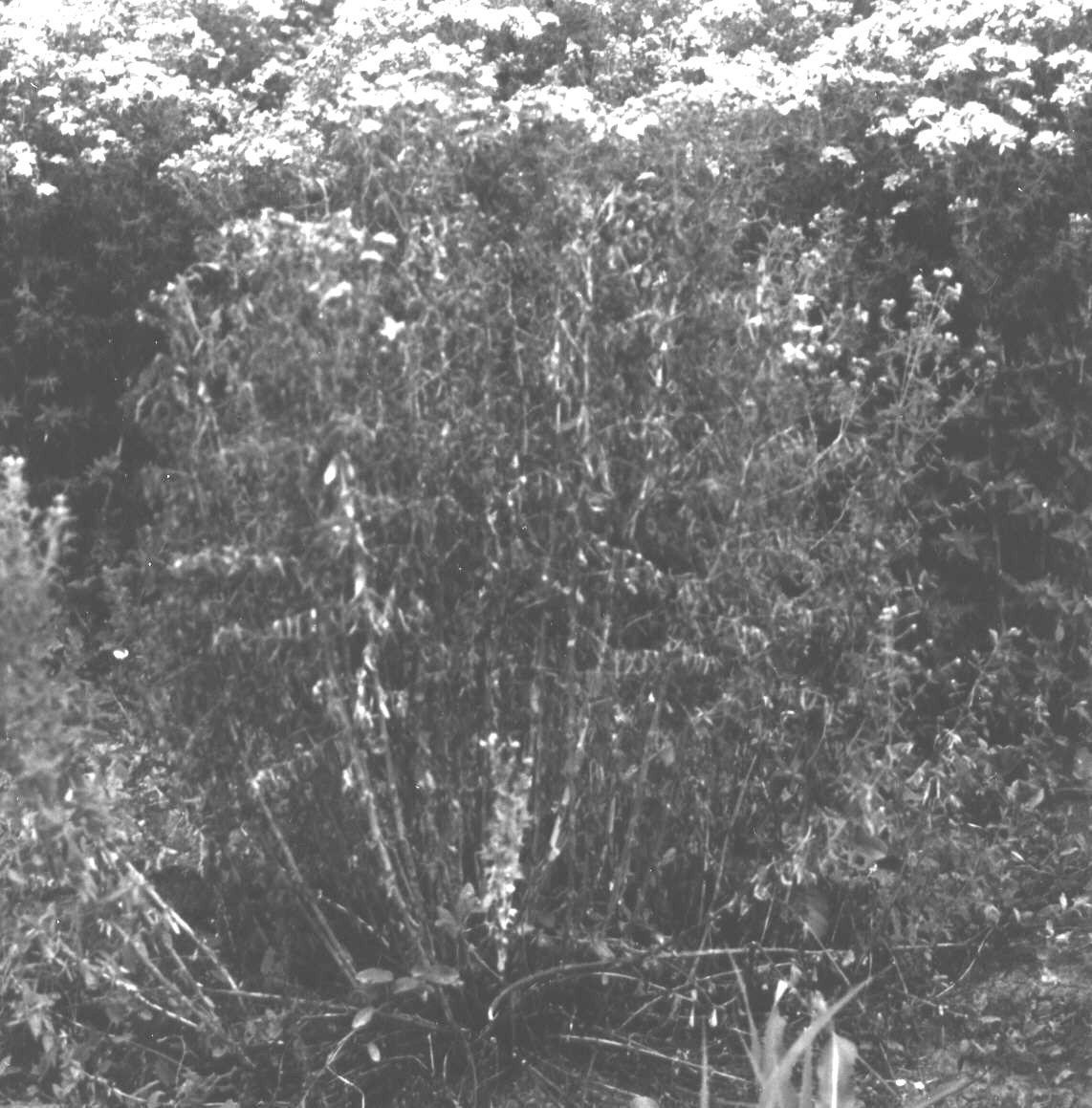
and death of some branches followed by the death of the whole plant (fig.2).
Some of the plants from one bush died out before flowering, some of them
produced flowers, but all bush died out after flower harvest.
|
|
|
|
|
|
Infected plant has brown and black discoloration of root and basal stem
(crown) crown and root rot; continuous stems and branches necrosis including
the inflorescence; black necrosis and desiccation of leaves and apics;
leaf distortion; and vascular discoloration. The disease was so severe
in 1999 that all plants of four plantations have died. In experimental
plants with different time of planting (fall and spring), the percentage
of dead plants reached 100%.
| Pathogenecity test |
|
Inoculated St. John's worth showed the same symptoms as in the field (wilting, necrosis and plant death) 6 weeks after inoculation. From inoculated plants, by reisolation the same colony as from the plant from the field has been isolated. The pathogen The fungus forms round colony with pink-colored aerial mycelia. Mycelia is coloring media pink. Micro and macro conidia are formed on mycelia and have dimensions 7.2 x 3.3 and 3.2 x 4.1 mm respectively. Hlamidospores are rounded and have dimension of 7.2 x 6.8 mm. |
 |
Effect of different media on colony growth of F. oxysporum have been studied by measuring colony diameter 8 days after growing on: PDA, WA, CMA, OMA, Czapek and MA. All five isolates have the similar growth on all (6) media. The best colony growth, after 8 days, have been achieved on OMA (colony diameter 77.86 mm), then followed by PDA and MA (colony diameter 77.40 mm and 69.53 mm respectively). CMA is least suitable media for colony growth of F. oxysporum (graph. 1).
Effect of temperature on colony growth of F. oxysporum has been
studied by growing the colony on PDA media at different temperatures. All
tested isolates have had the best growth at 25°C, (average colony diameter
after 8 days was 84.40 mm), followed by 30°C and 20°C (average colony diameter
after 8 days 72.83 and 70.13 mm respectively). The least growth of colony
was at 4°C (graph. 2).
|
|
|
 |
 |
Growing of 5 isolates of F. oxysporum on PDA media with different pH we have found that the best growth for all isolates have been when media has pH 8 (average colony diameter after 8 days was 85.55 mm) then on media pH 7 and pH 9 (colony diameter 85.20 mm and 84.60 mm, respectively, graph. 3).
So far, 64 formae speciales of F. oxysporum on different plants have been described (Watanabe, 1994). None were found on St.John's worth. We found F. oxysporum f. sp. fabae was found on faba beans in Serbia (Ivanović at al., 1987). There is no data of F. oxysporum on St. John's worth in literature.
Based on the results obtained in this research it may be concluded that the fungus isolated from diseased St. John's worth belongs to the Fusarium oxysporum Schl.
On artificially inoculated St. John's wort plants the symptoms obtained are very similar to those of naturally infected plants. Identification of the fungi has been done by morphology study and cultural characteristics of the pathogen. The fungus forms round colony with pink-colored aerial mycelia, micro- and macroconidia and hlamidospores. The best media for colony growth is oat mill agar. Optimal temperature for colony growth on PDA is 25°C, and the best colony growth was when pH of PDA media was 8.
Barl B.(2000): Universityof Saskatchewan, Saskatoon, Canada(personal communication).
Dhingra O.D. and Sinclair J.B. (1995): Basic Plant Pathology Methods (sec.eds.), CRC Press, Boca Raton, FL 355 pp.
Evstatieva Lj. (2000): Bulgarian Academy of Sciences, Institute of Botany, Sofia, Bulgaria (personal communication).
Gamliel A., Talmakatan Yunis H. and Katan J. (1996): Fusarium Wilt and Crown Rot of Sweet Basil: Involvement of Soil borne and Airborne Inoculums. Phytopathology, 86, 56-62.
Ivanović M., Ivanović D., Jovanović O. (1987): Fusarium oxysporum f. sp. fabae prouzrokovač truleži korena boba u Jugoslaviji. Zaštita bilja, 38 (4), br. 182, 373-380.
Radanović D., Jevdjović R. and Stepanović B. (2000): St. John's Wort (Hypericum perforatum L.) cultivation in South Banat region (Yugoslavia). First Conference on Medicinal and Aromatic Plants of Southern European Countries, Arandjelovac, Yugoslavia, May 29-June 3, 2000 (Book of Abstract).
Watanabe T. (1994): Pictorial Atlas of Soil and Seed Fungi- Morphologies of cultured fungi and key to species, pp. 411. Lewis Publishers, Washington DC, USA.