 |
 |
|
|
|
Studies of mycopopulation of medicinal plant seeds till this time were not conducted systematically. Remarkable yield reduction, as a cause of presence of different pathogens requires devotion of especial care to this problem. From this reason, the aims of our contribution were as follows:
Identification of the whole spectrum fungi present in seed samples;Tested St. John's wort seed samples were collected at four localities, i.e. Pančevo, Svrljig, Kačarevo and Gorobilje, during the years 1998 and 1999.
Determination of percents of abundance of these fungi;
Testing of pathogenicity of the most harmful species determined.
Health status of seed samples was evaluated by the means of standard methods (ISTA). This way, following fungi were determined: Fusarium oxysporum, Fusarium solani, Fusarium spp., Verticillium spp., Alternaria spp., Acremoniella atra, Aspergillus flavus, Penicillium spp., and Mucor spp. Among samples investigated, the most abundant species recorded on the contaminated seeds was Alternaria spp., followed with fungi belonging to the genus Fusarium.
St. John's wort seeds are populated with a number of microorganisms,
among which few of them could significantly decrease yield of herb.
Keywords: St. John's wort, seed, microflora.
Medicinal plants are very precious treasures of the nature. They have an invaluable economical importance for medical treatment, food, trade and export. As our country is very rich with the medicinal and aromatic plants, their exploitation is possible, but, at the same time, it is necessary to protect them against an uncontrolled use. Introducing the technology of cultivating the most jeopardized and rarest species that are important for the pharmaceutical industry on the growing places is one of the prerequisites for protecting medicinal plants in the nature.
For the last few years the world market has become more interested in St. John's wort seeds (Hypericum perforatum L.). Therefore the interest for it has increased, as well. Until 1997, all the necessary quantities of St. John's wort seeds in the region of Serbia were obtained only by collecting. However, in 1997. Institute for Medicinal Plants Research started with an experimental production of cultivating St. John's wort seeds. In this way growing of the seeds on the plantations is stimulated, and the pressure on populations in the nature is reduced.
At the very beginning, during the first years of experimental cultivating, we faced with the problems such as St. John's wort seeds' pathology and the rot of the most plants in the experimental field.
Therefore, we have started examining the health status of seed samples. Seed is the most important part of the plant production and both, the quality and the level of plant production depend on it. Even in the far away past the farm-producers found out how important it was for the yield. Lots of diseases are maintained and carried out from one vegetation to the other through the sowing of diseased seeds.
Diseased seeds enrich the parasite potential in the soil. The purpose of the phytopatological analysis of seed is to determine the sort of microorganisms in seeds. On the basis of these analyses we have come to the conclusion about the use value of the examined seed samples.
Seed samples from four locations; i.e. Pančevo, Kačarevo, Svrljig and
Gorobilje were examined in 1998 and 1999.
Standard phytopathological method (International Rules for Seed Testing
- Ista 1966: 1999) was used in those examinations, assuming:
method of isolation (400 seeds from each seed portion disinfected with 1% solution of NaOCl (3 minutes), than washed in sterile water);
seed incubation on filter papers (100 seeds in Petri dishes, diameter 25 cm);
seed incubation on nutritive ground (Potato dextrose Agar (PDA), Water Agar (WA), and Carnation Leaf-Piece Agar (CLA);
getting monosporial cultures;
determination and
pathogenicity.
St. John's wort seedlings raised in a greenhouse at temperatures of 25-30°C in the pots with sterile soil. The soil around the roots was diseased with fungus Fusarium oxysporum mixed with sterile water. One month later we isolated the same fungus in the necrotic parts of root and ground parts of stem. For determination we used the latest phytopatological literature.
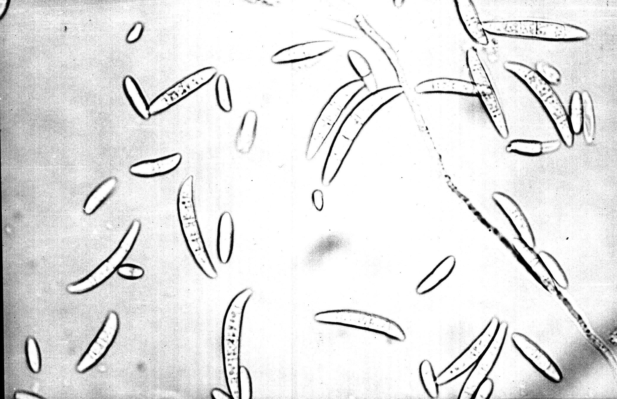
During the examinations of St. John's wort seeds there were some difficulties, as the seed is very small and it is not easy to handle with it (Figure 1). Besides, there were also some other difficulties because of the mixed diseases on the seed (several species of fungi on the same seed (Figure 2).
Ten species of fungi have been identified on the examined samples of St. John's wort seeds: Alternaria spp., Aspergillus spp., Fusarium oxysporum, Fusarium solani, Fusarium semitectum, Fusarium proliferatum, Penicillium spp., Epicoccum purpurascens, Verticillium spp. and Mucor spp. It could be seen that on St. John's wort seeds, fungi of the genus Fusarium appear most often.
|
|
|
|
|
|
| Locality | Pathogen % |
|
|
| Pančevo | Alternaria spp.
Fusarium spp. Aspergillus spp. Penicillium spp. Verticillium spp. |
6% 4% 3% 2% |
4% 4% 6% - |
| Kačarevo | Alternaria spp.
Fusarium spp. Aspergillus spp. Penicillium spp. Epicoccum purpurascens |
3% 3% 7% 2% |
4% 4% 5% - |
| Svrljig | Alternaria spp.
Fusarium spp. Aspergillus spp. Penicillium spp. |
6% 1% 3% |
4% 3% 3% |
| Gorobilje | Alternaria spp.
Fusarium spp. Aspergillus spp. Penicillium spp. Epicoccum purpurascens |
6% 1% 3% 2% |
6% 1% 3% 1,5% |
According to results presented in Table 1, it can be seen that the most frequent fungi, (expressed in percentages) belong to genus Alternaria and Fusarium. Fungi that belong to genus Penicillium, Aspergillus and Epicoccum were always present but in less occurrence. Differences between the seeds of the yield in 1998 and yield in 1999 were not obvious.
Fusarium oxysporum Schlecht. Emend Snyder & Hansen
This fungus was found both in all examined samples and in all locations. On the seed on filter paper the fungus forms sometimes rare and sometimes abundant aerial mycelium, white to pale violet, which changes the color of filter paper into pale violet (Figure 3). It is formed in most cultures within 4 days.
 |
 |
|
|
|
On the nutritive ground this fungus is forming a huge aerial mycelium of different colors, white, pale orange to pale violet (Figure 4) Aerial mycelium is fluffy, pale orange to pale violet. Fusarium oxysporum usually produces a pale to dark violet pigment in the agar, but some isolates do not produce any pigment.
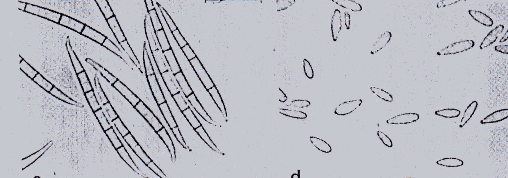
These fungi create micro and macroconidia and chlamydosphores on CLA
Agar. Microconidia, always present, are unicellular or bicellular, ellipsoidal,
oval, cylindrical 5.5-12 x 2-3.5 micron, formed abundantly in false heads
on short monophialides on hyphe.
The microconidia are hyaline non septate. Microconidia are formed from short monophialides or branched conidiophores in pale orange sporodohia (Figure 5). Macroconidia usually with 3 septates 33 x 4.1-27-42 x 3.0-4.5millimicron. Chlamydosphores, generally abundant in hyphe and conidia, terminal or intercalary "smooth-or-rough-walled" globose, subglobose single or in chains. |
 |
Fusarium oxysporum is one of the most variable species within the genus. It includes populations which cause vascular wilt diseases (Beckman, 1987), populations which cause root, crown, tuber, corm and bulb rots (Nelson et al., 1981) and populations which are soil saprophytes. Some members of Fusarium oxysporum also cause opportunistic infections of humans and other animals. (Rebbel, 1981.)
Fusarium oxysporum
In order to check if the examined fungus is pathogenic for St. John's wort seeds we made a trial in greenhouse in pots (20 cm in diameter) filled with sterile water. When the sterilization was over the soil was moistured with sterile water. The pure culture of the fungus, genus Fusarium oxysporum, was cultivated in Petri dishes. The cultures were 7 days old. With the culture of the fungus from one of the Petri dishes we infected the soil in one pot. We used a sterile scalpel to chop the rest of the ground mixed with the soil in the pot. In those pots we cultivated seedlings of St. John's wort seeds. There were 15 pots. The soil around the root systems in 10 pots was infected in the same way with the culture of the fungus. The other 5 pots were used for control. In those pots everything was done in the same way, but without the presence of the fungus. The pots were kept into greenhouse at the temperatures of 25°C - 30°C, (12 hours 25°C, 12 hours 30°C; air humidity 90%; 12 hours in light, 12 hours in dark, alternately).
The first symptoms appeared one month later, the leaves turned into yellow, and necrosis noticed on the lower parts of the stem and on the root system. (Figure 6). Reisolation - the clean culture Fusarium oxysporum was obtained by separating parasite fungus from the pathogenic plants from the infected pots and growing them on the nutritive ground on PDA. When the morphological characteristics were examined we came to the conclusion that the reisolated fungus was identical to the one we got before the trial. Control plants were healthy. (Figure 7).
 |
 |
|
|
|
Fusarium solani (Mart.) Appel & Wollenw. Emend. Snyder & Hansen
On the seed on filter paper it creates white, creamy and diluted mycelia. Pigmentation on filter paper varies, as in Fusarium oxysporum, from pale to dark violet.
 |
 |
|
|
|
On PDA it forms a rare aerial mycelia, at the beginning white, creamy, and at the end dark violet (Figure 8). Pigmentation is dark violet. They are formed in most cultures within a few days.
It forms microconidia, macroconidia and chlamydosphores. The microconidia are formed on long monophialides in false heads (Figure 9). They are one celled or two celled, hyaline, oval, ellipsoidal, cylindrical 7.5-16.2 x 2.2-4.0 micron.
The macroconidia are formed from long monophialides, on branched conidiophores, usually within 4-5 days. The macroconidia are usually with 3 or 5 septs, even though there are the macroconidia with 7 septs. Dimensions vary depending on isolates, 36-58 x 4-6.5 micron. The chlamidosphores - more or less abundant, terminal or intercalary in hyphae and conidia, globose to subglobose, single in pairs, chains or clusters. Chlamidosphores are formed in most cultures within 2 to 3 weeks.
Fusarium semitectum Berk. & Rav.
A white coating is formed both on germinated and non-germinated seeds after incubation in a humid chamber, at the temperature of 25°C. On the seeds, on nutritive ground, a white, very abundant aerial mycelium is formed, too. Colonies grow very quickly up to 4cm within 3 days at the temperature of 25°C on PDA (Figure 10).
Pigmentation - color of a peach (whitish, flesh, peach, gradually changing to ochraceous, avellaneous and finally buff-brown).
 |
 |
|
|
|
Only macroconidia are present (Figure 11), usually 4-5 septate measuring: (24-45 x 4.5-6.1) conidia, rather thick-walled, predominantly straight and spindle-shaped to lanceolate, sometimes slightly curved and falcate, gradually tapering toward each end with a hooked apical and mostly indistinct conical or wedge-shaped, apedicellate, sometimes apiculate basal cell. Conidia are formed from polyohialides on the aerial hyphae.
Chlamydospore formation is variable, often sparse, mostly intercallary. Fusariumsemitectum is isolated from the sage seeds (Salvia officinalis) and peppermint stolones.
Fusarium proliferatum (Matsushima) Nirenberg
At seed they form floccose mycelia, dirty white in color that later become light orange, like color of pear. At growing media the colony grows fast.
Colonies are fast growing, at 25°C on PDA within 3 days up to 4 cm in diameter; some strains, however are only moderately fast growing.
Aerial mycelium whites floccose, which may become grayish violet or grayish magenta with age (Figure 12).
 |
 |
|
|
|
Pigmentation in the agar is quite variable, ranging from no pigmentation or grayish orange in some isolates to violet gray, dark violet, almost black in others.
Sporulation starting after 3 days as microconidia in on CLA. Microconidia are formed abundantly in chains from polyphialides, which may proliferate, less often from monophialides. Microconidia also form false heads. The microconidia always clavate, usually 1or 2 septate, measuring: 8.8 x 3.0 mostly 8-11 x 2.6-3.7m .
Macroconidia are produced in pale orange sporodochia, which may be obscured
by the mycelium. Macroconidia are long, slender, falcate to almost straight,
usually 3-5 septate and thin-walled, measuring: 33 x 3.0 mostly 24-58 x
2.7-3.8m (Figure 13).
Chlamydospores are absent.
Our research work has proved that St. John's wort seeds were exposed to the attacks of numerous parasite and saprophyte fungi. Parasite fungi cause the seed pathology during the vegetation. Some parasite potentials of the fungus pathogens are enriched with the infected seed sowing. Our research found out 4 species of the genus Fusarium. According to the data from Russian and Bulgarian literature only Fusarium spp. was found on St. John's wort seeds.
The species of the genus Alternaria spp. are present most of all in our research work. Health status of the seed and its use power depend on the kind and number of microorganisms settled on the surface or interior parts of the seeds.
On the basis of the obtained results the following could be concluded:
10 species of the fungi, of the genus Fusarium mostly, were isolated from St. John's wort seeds;
The mixed infection of several species of fungi was found on St. John's wort seeds;
We determined and described 4 species of the genus Fusarium: Fusarium oxysporum, Fusarium solani, Fusarium semitectum and Fusarium proliferatum;
We examined the pathogenicity of the fungus Fusarium oxysporum, and we came to the conclusion that this fungus causes decay of St. John's wort seeds in growing places;
The species of the genus Alternaria are found most of all;
The presence of so-called Ware-house fungi, such as Aspergillus, Penicillium, Mucor was registered; these fungi haven't been described on St. John's wort seeds so far.
Bilai V.I. (1955): Fusarium, Kiev, Ukr.SSR Acid Sci. 282, 286.
Beckman C.H.(1987): The nature of wilt diseases of plants, APS Press, Minnesota.
Boot C. (1971): The genus Fusarium. Commonwealth Mycological Institute Kew, Surrey, England.72, 76, 116, 130-134.
Gerlach&Nirenberg (1982): The genus Fusarium a pictorial atlas, Mitteilungen aus der Biologischen Bundesanstalt fur Land- und Forstwirschaft. Berlin-Dahlem., 155-159, 309-316, 345-350.
Dobrozrakova T.L., Letova M.F., Stepanov K.M., Hohrjakov M.K. (1956): Opredelitel nabolestite porasteliayta, Moscow-Leningrad, Nauka. 246-247.
Joffe A.Z. (1986): Fusarium species: Their biology and toxicology. John Wiley and Sons, New York.
Lester W. Burgess at al. (1994): Laboratory manual for Fusarium research, Sydney, 62, 74, 76, 116.
Malone J.P., Muskett A.E. (1964): Seed borne fungi. Plant Pathology Division, Ministry of Agriculture, The Queen's University, Belfast.
Nelson P.E., Toussoun T.A. & Cook R.J., Eds. (1981): Fusarium: Disseases, Biology and Taxonnomy, The Pennsylvania State University Press, University Park and London.