 |
 |
|
|
|
Stolone-born fungi are one of the most important causes of decrease of the yield in the cultivation of peppermint. From this reason, the aim of our studies was isolation and determination of pathogenic microorganisms from mint stolones. Stolones of introduced cultivar Mitcham were analyzed. Among seven species of microorganisms determined, the most important are species belonging to the genus Fusarium and Verticillium. In samples, collected from the experimental field of Institute for Medicinal Plant Research located in the neighborhood of Pančevo, following species were isolated and determined.
Fusarium culmorum (W.G. Smith) Sacc.Results of our investigations confirmed that mint stolones are populated with pathogen type micro-organisms‚ as well as those of saprophyte nature. Fungi of the genus Verticillium and Fusarium causes specific chloroses and necroses characteristic for these phytopathogens.
Fusarim equiseti (Corda) Sacc.
Fusarim smitectum Berk & Rav.
Fusarim subglutinans Wr. Reink.
Verticillium lateritum Fr.
Verticillium dahliae Klebhan&Smith
Penicillum spp.
Trichoteciun roseum Link & Fries
Alternaria spp.
Keywords: mint, micro-organisms, fungi, pathogens, Fusarium, Verticillium.
A large number of pathogenic microorganisms, especially fungi, has been recorded on this plant. In our conditions of mint growing Puccinia menthae Pers., causes the most important yield loses and the most serious problems to the producers. In the last few years the biggest problems in mint growing are caused by the fungi of the genus Fusarium and Verticillium. According to the results from the Russian literature, other important phytopathogens are Fusarium spp., then Septoria menthae Oud., Septoria menthicola Sacc., Ramularia menthicola Sacc., Erysiphe cichoracearum DC. f. menthe. Jacz. and Peronospora stigmaticola Raunk.
Significant yield reduction and low quality of drug, as a result of activity of different fungi, requires more attention to be devoted to this problem. The leaves attacked by those fungi could not be sold or used for further production, as besides a bad quality of drug and reduced quantities of essential oils, there is still a danger of micotoxine produced by the fungi.
The samples that are examined in this research are taken from the experimental field of Institute for Medicinal Plant Research in Pančevo in the period from 1997 to 2000. The analysis of the health status of the mint stolones is done with the standard phytopatological methods such as:
Isolation of microorganisms from stolones.
Obtaining of monosporial cultures.
Morphological characteristics of the obtained isolates: the rate of colony growth, the nature of aerial mycelium, the presence of pigmentation, the look of conidiophores, the look of conidia, the way of conidia formation, production of chlamydosphores, picnide or sometimes sclerotia and strome formation. At least one hundred conidia of monosphorial culture were measured.
Determination on the basis of morphological, biometric and cultivating characteristics of the examined fungi, taking into consideration the symptoms of disease if they were typical. Both classic and recent literature have been studied during determination.
It was identified 9 kinds of fungi on the examined stolones. The frequency of disease was determined as presented in Table 1.
| Fusarium semitectum Berk Rav. |
|
| Fusarium subglutinans Wr. Reink |
|
| Fusarium culmorum (W.G.Smith) Sacc. |
|
| Fusarium equiseti (Corda )Sacc. |
|
| Fusarium proliferatum (Matsushima) Nir |
|
| Verticillium dahliae Klebahn, Smith |
|
| Verticillium lateritium (Fr.) |
|
| Alternaria spp. |
|
| Penicillium spp. |
|
The most significant fungi that could cause a serious economic damage on the peppermint are of the genus Fusarium and Verticillium. The damage could be caused even by some other species, so called saprophyte fungi, however, they were not so significant (Alternaria spp., Penicillium spp., Aspergillus spp., Trichothecium roseum and others).
The damage depends mostly on the conditions for parasite development, on species, mechanical damage and keeping conditions.
Fusarium culmorum (W.G.Smith) Sacc.
It has been found in most examined species, especially in cultivar Mitcham. The stolones infected with this fungus have a characteristic appearance. There is a plenty of mycelia on them, changing the color from white and yellow to pink, sporulating abundantly.
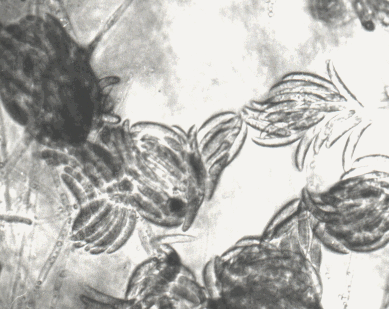
Colonies grow very quickly, 5.5 cm in diameter within 3 days, on PDA medium, at the temperature of 25°C. Aerial mycelia, abundant, changing the color from white, yellow to pink and finally becoming dark red in the middle of the box and white at the edge (Figure 1).
|
|
|
|
|
|
Pigmentation is dark red with dipped hyphae.
Sporulation is very fast, macroconidia in aerial mycelium are formed
within 7 days, whereas sporodohia, full of macroconidia, are formed at
the bottom of a glass, in older cultures. Microconidia are absent.
Conidiosphores are branched with phyalides by side, monophyalides only.
Macroconidia are typical for this species with thick walls, septate,
different shaped, curved or straight, wide to the point, tapering towards
the basal part, or they could be wide in the middle, tapering towards the
ends (Figure 2). There are plenty of chlamydosphores.
Fusarium subglutinans (Wollenw.&Reinking) Nelson.
On stolones Fusarium subglutinans is producing white aerial mycelium that is usually mixed with some species of other genus (Figure 3).
 |
 |
|
|
|
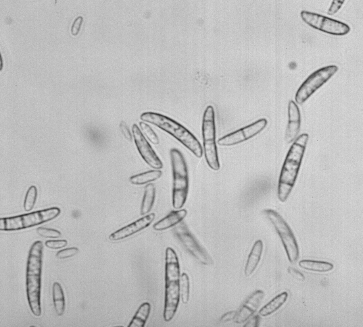
The cultures are similar in Fusarium monoliforme. On nutritive ground this fungus forms floccose, white to greyish-white mycelia, changing the colour to vinaceous purple, becoming purple to dark purple. Macroconidia and microconidia are present. Chlamydosphores are absent. Microconidia appear within 2-3 days. Microconidia are aseptate, hyaline, oval to obclavate, 8-11.8 x 2.5-3 m. Macroconidia are usually produced in abundance in false-heads mainly from polyphialides but also from monophialides.Some isolates also produce longer microconidia which are spindle-shaped, 2 to 3 septate and which may be apiculate at each end and appear to be similar to the macroconidia.
Macroconidia are formed in pale orange sporodochia. They are thin-walled, falcate, long, 3 to 5 septate, 33-53 x 3-4.5m. The apical cell is usually curved and tapered to the point. The basal cell is foot-shaped (Figure 4).
Fusarium equiseti (Corda) Sacc.
We have isolated this species of the genus Fusarium from mint stolones. It produces abundant dirty white mycelium on infected stolones.
This fungus grows very quickly, 4cm at the temperature of 25°C on PDA, on nutritive ground. Aerial mycelium is white and abundant. Pigmentation is dark brown. Macroconidia are present, produced abundantly in orange sporodochia. Macroconidia are usually 5 to 7 septate, measuring: 35-60 X 3.5-5.9 micron, with a tapering apical cell and a distinct foot-shaped base to the basal cell. (Figure 5). Macroconidia are produced from monophialides on branched conidiophores in sporodochia.
Chlamydosphores intercalary, solitary, in chains or in knots, globose, 7 to 9 micron in diameter (Figure 6). Fusarium equiseti is a cosmopolitan in soil. According to the data from literature it is an important pathogen.
 |
 |
|
|
|
Fusarium prolifratum (Matsushima) Nir
An abundant dirty-white mycelium is formed on infected stolones. The colour of stolones is changing from grey to dark grey, from brown and red to dark brown. From these stolones the fungus of the genus Fusarium prolifatum was isolated and determined. It was found on St. John's wort seeds. The fungus was described in detail.
Fusarium semitectum Berk Rav.
This fungus was isolated from stolones of all the examined samples. The greatest number of isolates was isolated from the mint stolones. During the examination of the health status of seed, stolones were covered with dirty white mycelium. Following microscoping the presence of fungus of the genus Fusarium, later determined as Fusarium semitectum Berk Rav., was found out.
Verticillium dahliae Klebhan, Smith
A large number of isolates determined as Verticillium dahliae was isolated from the samples of infected stolones and the root systems showing the wilt symptoms. The wilting comes after chlorosa of leaves falling down in lower parts.
Later, in the next phase of the disease development, the whole plant gets withered. After microscoping the cross section of stolones and the ground parts of the stem, the necrosis of vascular tissue, like a dark color of diseased tissue, was discovered. The necrosis of vascular tissue is a characteristic diagnostic symptom, as it appears in the zone of carrying out elements of diseased plants, widening from the base towards the top. The long, narrow and wide stripes on the cross section show the necrosis process. It was found a lot of micro sclerotium (Figure 7).
 |
 |
|
|
|
Isolation of the fungus on nutritive ground on PDA is very simple. Within 3-4 days a white colony is developed around the fragments of diseased parts of the tissue. Conidiophores are transparent, branched aside. Conidia are one-celled, hyaline, oval formed in groups, like a small head, measuring: 3.1-4.9 x 4.9-7.3 m.
Symptoms are manifested through leaf chlorosis, followed by necrosis starting from the base to the plant tops (Figure 8). Finally, wilting symptoms resulting in appearance of empty rows that could be easily recorded.
The results, obtained during this investigation proved that stolones have a very significant role in mint pathology. Because of that and in order to have healthy seedlings as well as less damage that could be caused by this fungus, it is very important to have seedlings of good quality.
From the samples of diseased stolones and ground parts of the stem, 9 species of different cultivars were isolated. In this investigation 5 species of the genus Fusarium and 2 species of the genus Verticillium were isolated.
Species of the genus Fusarium haven't been described on this plant so far. According to the data from the literature, only Fusarium spp., was isolated on mint (Dobrozrakova, 1956) and (Hristov, 1972).
On the basis of the results in examining the presence of microflora on mint stolones, in 1998. And 1999., when several methods were applied, it could be concluded as follows.
The presence of 9 species of fungi was found out.
The fungi were of different cultivars.
The fungi of the genus Fusarium (F. semitectum, F. subglutinans, F. culmorum, F. equiseti and F. proliferatum) are found most of all.
Species of the genus Verticillium are found as well, causing withering of plants in mint growing. During the last few years they cause some serious problems in the production of this culture.
Beside these species it has been isolated and determined saprophyte species of the genus Penicillium and Alternaria, which were present on the mint stolones, causing no damage at all.
Bilai V.I. (1955): Fusarium, Kiev, Ukr. SSR Acad. Sci. 259, 283, 299.
Boot C. (1971): The genus Fusarium, Commonwealth Mycological Institute Kew, Surrey, England, 94-96, 127-129, 157-159, 173-176.
Gerlach&Nirenberg (1982): The genus Fusarium a pictorial atlas. Mitteilungen aus der Biollogischen Bundesanstalt fur Land-und Forstwirtchaft. Berlin-Dahlem, 155-159, 177-180, 225-228, 309-316.
Dobrozrakova T.L., Letova M.F., Stepanov K.M., Hohrjakov M.K. (1956): Opredelitel nabolestite porasteliayta. Moscow-Leningrad, Nauka, 245-246.
Joffe A.Z. (1986): Fusarium species: Their biology and toxicology. John Wiley and Sons, New York.
Kojič M., Jančić R. eds. (1998): Pitoma nana (Mentha x piperita L.) i druge vrste roda Mentha L.- Institut za proučavanje lekovitog bilja "Dr Josif Pančić" i IP "Ecolibri", Beograd.
Lester W. Burgess (1994): Laboratory manual for Fusarium research, Sydney 62, 65, 82, 109, 118.
Pidopličko N.M. (1977): Gribi-paraziti kulturnih rastenii, Kiev, Tom 2, 55-60, 82-83.
Malone J.P., Muskett A.E. (1964): Seed borne fungi, Plant Pathology Division, Ministry of Agriculture, The Queen's University, Belfast, p.179-300.